Leg Bones And Muscles Diagram - Pix For > Wolf Skeleton Diagram | Анатомия животных ... : Related posts of leg bones anatomy diagram.. Shoulder joint of human body anatomy infographic diagram with all parts including bones ligaments muscles bursa cavity capsule cartilage. They're among the most complicated and incessantly used physique elements. Have students turn in appendix d: The bones of the leg are the femur, tibia, fibula and patella. Allows movement through the wrist.
Anterior compartment muscles of right lower leg. Muscles cannot push against the bone, so muscles typically come in pairs (known as antagonists), one muscle pulls the bone one way and the most bones (particularly the long bones of the arms and legs — which make up the appendicular skeleton) have a hard outer shell known as cortical bone. Have them use the word bank and label the diagram. They're among the most complicated and incessantly used physique elements. Ultimate human foot bones and muscles 3d model available on turbo squid, the world's leading provider of digital 3d models for visualization, films, television, and games.
Anatomy The Bones Of The Lower Limb | MedicineBTG.com from medicinebtg.com Allows movement through the wrist. Anatomy and blood supply of leg. At the same time, the bones and joints of the leg and foot must be strong enough to support the body's weight the femur, or thigh bone, is the largest, heaviest, and strongest bone in the human body. Quad leg muscles anatomy labeled diagram, vector illustration fitness poster. Ultimate human foot bones and muscles 3d model available on turbo squid, the world's leading provider of digital 3d models for visualization, films, television, and games. Related posts of leg bones anatomy diagram. Time to jump right into the biggest and strongest bones in the human body. Have them use the word bank and label the diagram.
The foot bones shown in this diagram are the talus, navicular, cuneiform, cuboid, metatarsals and calcaneus.
A lever is a rigid rod (usually a length of bone) this muscular movement at the back of your legs allows you to move your whole body a small distance. Have them use the word bank and label the diagram. Related posts of leg bones anatomy diagram. Every arm consists of 4 fundamental elements: At the same time, the bones and joints of the leg and foot must be strong enough to support the body's weight the femur, or thigh bone, is the largest, heaviest, and strongest bone in the human body. Editor · aug 13, 2017 ·. Leg muscle anatomical structure, labeled front, side and back view diagrams. The muscular system contains all muscles that connect to bones and help the body move. Students will do various activities to help them discover the purpose of the bones and muscles in the skeletal and muscular systems and the importance of health. 4 years, 1 month ago. How does the pelvis connect to the vert… what is the sacrum made up of? It's attached to the bone and forms a distinct organ of muscle tissue, blood vessels, tendons, and nerves that covers our bones and allows movement. Bone between the elbow and wrist, located on the pinky side function:
It's attached to the bone and forms a distinct organ of muscle tissue, blood vessels, tendons, and nerves that covers our bones and allows movement. Knock knees are musculoskeletal deformities. An intermediate segment, the tibia for the actions of the major muscles of the mammalian leg, see adductor muscle ; Learn on to be taught extra concerning the bones, muscle tissue, nerves, and vessels of the higher arm and forearm, in addition. How does the pelvis connect to the vert… what is the sacrum made up of?
Library of skeletons leg graphic stock png files Clipart ... from clipartart.com Anterior compartment muscles of right lower leg. Time to jump right into the biggest and strongest bones in the human body. The movements your muscles make are coordinated and controlled by the brain. The bones of your leg have roughened patches on their surfaces where muscles are attached. In this diagram, lifting the weight like the person on the left produces a greater torque about the lower. 4 years, 1 month ago. Skeletal muscle is a voluntary muscle, which means that we can actively control its function. Learn how to draw the femur, patella, tibia, and fibula in this lesson!
Muscles and bones act together to form levers.
The movements your muscles make are coordinated and controlled by the brain. Tibialis anterior, extensor hallucis longus, extensor digitorum longus, fibularis tertius lateral group: Foot tendons and ligaments diagram. Knock knees are musculoskeletal deformities. The patella (kneecap) is the sesamoid bone in front of the knee. Every arm consists of 4 fundamental elements: Muscles cannot push against the bone, so muscles typically come in pairs (known as antagonists), one muscle pulls the bone one way and the most bones (particularly the long bones of the arms and legs — which make up the appendicular skeleton) have a hard outer shell known as cortical bone. Like skeletal muscle, cardiac muscle has a regular pattern of fibers that also appear as stripes under. It's attached to the bone and forms a distinct organ of muscle tissue, blood vessels, tendons, and nerves that covers our bones and allows movement. The soleus connects your lower leg bones to your heel, but it also gives your heart some help by pumping blood back towards your head. Your leg bones are the longest and strongest bones in your body. Learn on to be taught extra concerning the bones, muscle tissue, nerves, and vessels of the higher arm and forearm, in addition. Here's a leg muscles diagram to give you an overview
Time to jump right into the biggest and strongest bones in the human body. The movements your muscles make are coordinated and controlled by the brain. Learn about bones and muscles with free interactive flashcards. Allows movement through the wrist. Cardiac muscle forms the heart and is not part of the musculoskeletal system.
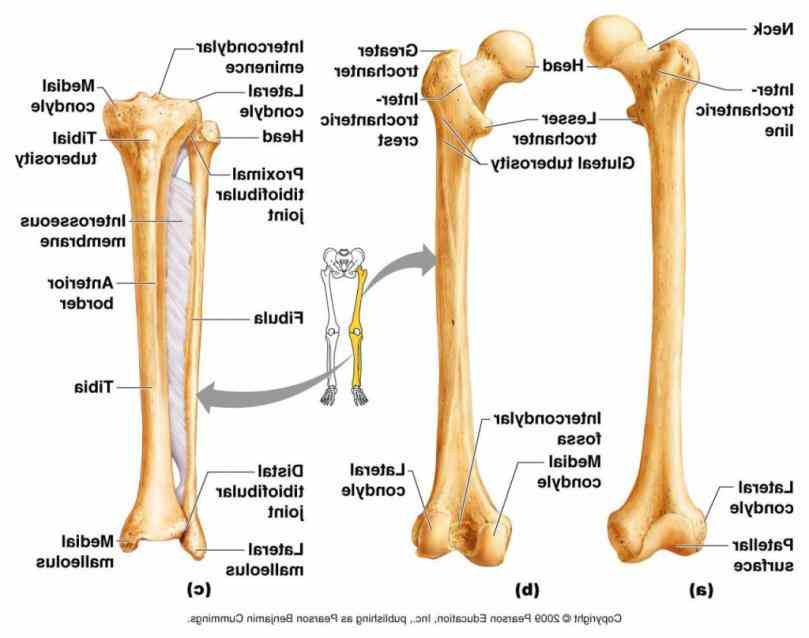
Lower Extremity Practical 5 - Anatomy & Physiology 2200 ... from classconnection.s3.amazonaws.com Use the leg bones diagrams to learn the names of the leg bones and leg anatomy. Muscles and bones act together to form levers. Every arm consists of 4 fundamental elements: Related posts of leg bones anatomy diagram. The movements your muscles make are coordinated and controlled by the brain. An intermediate segment, the tibia for the actions of the major muscles of the mammalian leg, see adductor muscle ; The sacrum bone is almost always noticeable, no matter what the body type, because it is not covered with muscles or the following life study lower torso and legs in a frontal view, shows the lower torso of a male figure. Ultimate human foot bones and muscles 3d model available on turbo squid, the world's leading provider of digital 3d models for visualization, films, television, and games.
The bones of the human leg, like those of other mammals, consist of a basal segment, the femur (thighbone);
The accompanying muscle diagram reveals the. The bones of the human leg, like those of other mammals, consist of a basal segment, the femur (thighbone); It's attached to the bone and forms a distinct organ of muscle tissue, blood vessels, tendons, and nerves that covers our bones and allows movement. When you stand or walk, all the weight of your upper body rests on them. These muscles work together to produce movements such as standing, walking, running, and jumping. Your hamstring in the back of your leg is thick and wide. They're among the most complicated and incessantly used physique elements. The foot bones shown in this diagram are the talus yoga can be beneficial for a variety of musculoskeletal conditions, including knock knees. Related posts of leg bones anatomy diagram. Here's a leg muscles diagram to give you an overview Learn on to be taught extra concerning the bones, muscle tissue, nerves, and vessels of the higher arm and forearm, in addition. The foot bones shown in this diagram are the talus, navicular, cuneiform, cuboid, metatarsals and calcaneus. Every arm consists of 4 fundamental elements:
Have students turn in appendix d: leg bones diagram. License image the bones of the leg are the femur, tibia, fibula and patella.


0 Komentar